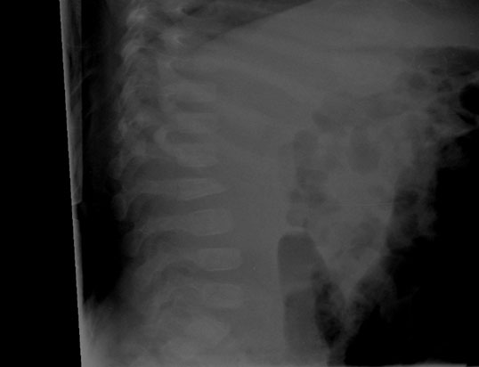
The lateral spine X-ray of a seven-week-old infant is below. The baby presented with multiple rib fractures, retinal hemorrhages, subdural hemorrhages, and a left distal radial fracture.

Accessible Version
A lateral radiograph of an infant’s thorax with lumbar vertebrae collapse at the anterior edge.
Which of the following is true?
◯
A. This finding shows a compression fracture of L2.
◯
B. This injury would be best observed using radionuclide scintigraphy.
◯
C. This injury is reportedly an uncommon finding in child abuse.
◯
D. Both A and C.
Which of the following is true?
◯
A. This finding shows a compression fracture of L2.
◯
B. This injury would be best observed using radionuclide scintigraphy.
◯
C. This injury is reportedly an uncommon finding in child abuse.
✓
D. Both A and C.
The best answer is D
Injuries can occur at any place along the spine but usually occur at the thoracolumbar junction. There are three histologic/radiologic patterns of vertebral injury described by Kleinman:
- Mild compression deformity with intact end-plates, as seen in this patient's X-ray (L2), usually from axial loading.
- Fractures of the anterior-superior end-plate of the vertebral body without loss of gross height of the vertebral body. It may result in irregularities of the vertebral body or discrete bone fragments, which usually are due to a hyperflexion injury. End-plate injury may result in significant growth disturbance and persistent vertebral deformity.
- Both compression deformity and superior end-plate extension.
Severe hyperflexion may result in disk rupture and herniation. If there is disruption of the end-plate or signs of neurologic damage, a spine MRI should be obtained. Severe hyperflexion may also result in injury to the posterior vertebral elements, usually the spinous processes.
Bone scan is insensitive for fractures of the vertebral body, as well as Classic Metaphyseal Lesions (CML) and skull fractures, but may be useful for demonstration of fractures of the transverse and spinous processes.
Most likely, vertebral fractures are more common than are reported (0-3%). They are probably missed because they are commonly asymptomatic in infants.
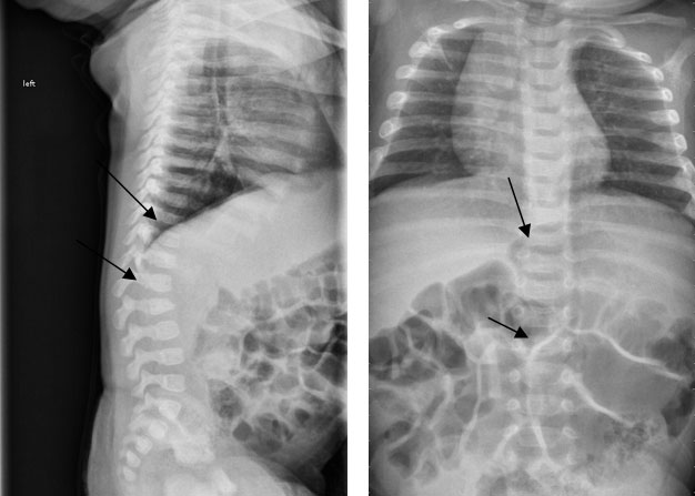
Note in the X-ray below from the follow-up bone survey that there is not only an L2 compression fracture, as seen on the initial lateral X-ray of the spine, but there is also a T10 compression as well as multiple rib fractures (T6-10 on the left and T4-7 on the right).
Complications of these injuries include nerve damage, which may present with flaccid paralysis, and long-term complications, such as nerve damage, spine deformities and scoliosis.

- Kleinman PK, ed. Diagnostic Imaging of Child Abuse, 3rd Ed. Cambridge University Press, Cambridge, United Kingdom. 2015.


